


1. Introduction
The nervous system serves as the primary controlling, regulatory, and communicating network within the human body. It acts as the central hub for all higher cognitive capabilities, including thought processes, learning, and memory consolidation.
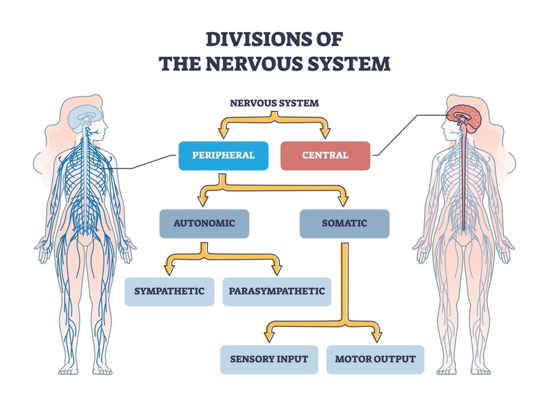
From an anatomical perspective, the nervous system is categorized into two interconnected structural components:
- The Central Nervous System (CNS): The primary integration and command center.
- The Peripheral Nervous System (PNS): The conduit for communication between the CNS and the rest of the body.
2. Anatomical Divisions
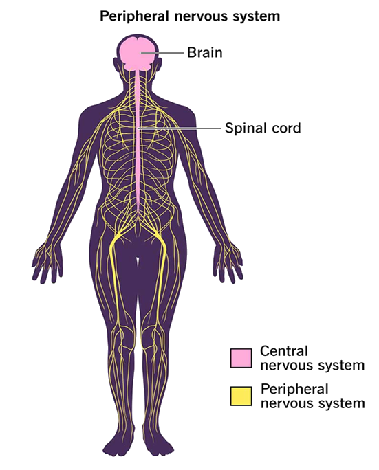
A. The Central Nervous System (CNS)
The CNS is anatomically composed of two main structures: the brain and the spinal cord.
- Functional Significance: It is designated as "central" because it integrates all incoming sensory information, coordinates internal homeostatic mechanisms, and influences the physiological activities of all bodily systems.
B. The Peripheral Nervous System (PNS)
The PNS comprises all neural structures located outside the boundaries of the brain and spinal cord, specifically the cranial/spinal nerves and their associated ganglia.
- Functional Significance: The primary functional role of the PNS is to establish a bidirectional communication pathway, tethering the central nervous system (CNS) to the peripheral limbs, organs, and tissues.
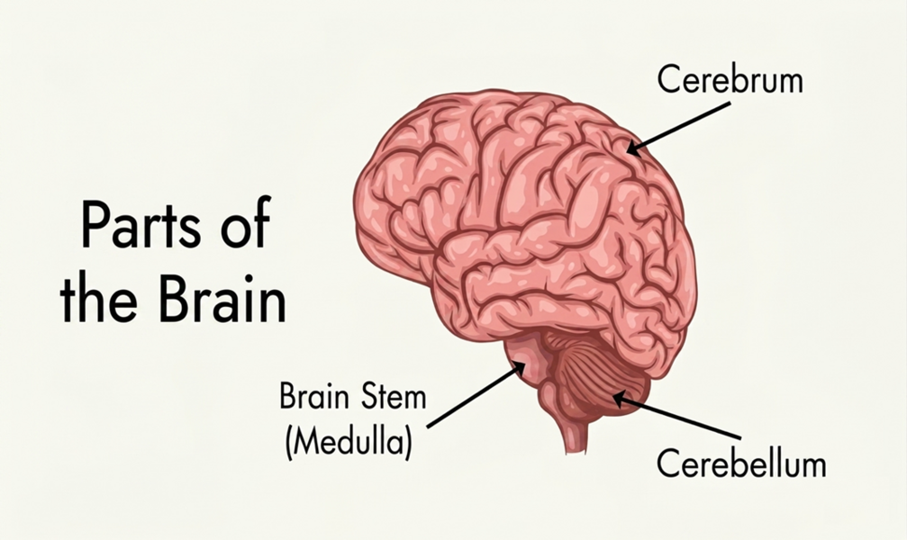
3. Major Anatomical Regions of the Brain
The encephalon (brain) is structurally subdivided into major regions, each executing distinct physiological and regulatory tasks:
- A. Cerebrum: As the largest region of the brain, it is primarily responsible for complex cognitive functions, including thought processing, memory retention, emotional perception, and the integration of somatic sensory modalities.
- B. Cerebellum: Positioned inferior to the cerebrum, this structure is the principal regulator of motor coordination, posture, and the maintenance of somatic balance.
- C. Brainstem: Positioned at the base of the brain, it is responsible for vital autonomic regulations, including the cardiac cycle (heartbeat), respiratory rhythm, arterial blood pressure, and the sleep-wake cycle.
4. Cellular Composition of the Nervous System
The histology of nervous tissue exhibits two major populations of specialized cells:
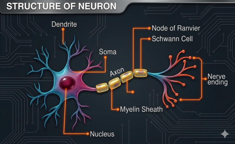
A. Neurons (Nerve Cells)
The neuron represents the fundamental structural and functional unit of the nervous system. Structurally, a classic neuron is comprised of a cell body (soma) and elongated cytoplasmic extensions specialized for conducting bioelectric impulses across vast distances. Neurons propagate signals to target cells through these specialized, thin fibers known as axons.
Cytological Components of a Neuron:
- Cell Body (Soma): The metabolic core of the cell, housing the nucleus along with critical intracellular organelles required for cellular maintenance (e.g., mitochondria).
- Dendrites: Branch-like projections specialized in receiving incoming neural impulses and propagating them centripetally toward the soma.
- Axon: A singular, elongated, and thin cytoplasmic process specialized in conducting action potentials away from the soma toward adjacent neurons or effector tissues.
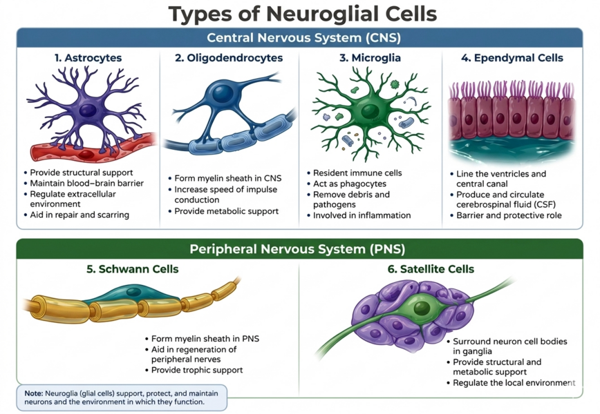
B. Neuroglia (Glial Cells)
Glial cells constitute the supportive framework of nervous tissue. Rather than conducting action potentials, their primary physiological responsibilities include maintaining extracellular homeostasis, synthesizing the insulating myelin sheath, and providing structural support and immunological protection for neurons.
- Prominent Examples: Astrocytes, Oligodendrocytes, and Microglia.
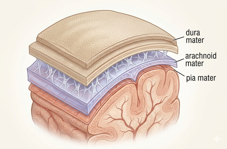
5. Protective Coverings: The Spinal Cord and Meninges
The spinal cord is structurally enveloped and protected by three distinct connective tissue layers known collectively as the Meninges:
- Dura Mater: The thick, dense, and highly fibrous outermost protective membrane.
- Arachnoid Mater: The thinner, avascular middle layer exhibiting a spiderweb-like architecture.
- Pia Mater: The delicate, highly vascularized innermost layer that directly adheres to the surface of the neural tissue.
الترجمة
الجهاز العصبي البشري: نظرة عامة على البنية والوظيفة
1. مقدمة (Introduction)
يُعد الجهاز العصبي شبكة التحكم، والتنظيم، والتواصل الرئيسية في الجسم البشري؛ إذ يمثل المركز الأساسي لجميع الأنشطة المعرفية والعقلية العليا، بما في ذلك التفكير، والتعلم، وترسيخ الذاكرة.
ومن الناحية التشريحية، يُصنف الجهاز العصبي إلى مكونين بنيويين مترابطين:
- الجهاز العصبي المركزي (CNS): ويمثل مركز التكامل والتحكم الرئيسي في الجسم.
- الجهاز العصبي المحيطي (PNS): ويمثل ممر التواصل بين الجهاز العصبي المركزي وبقية أعضاء الجسم.
2. الأقسام التشريحية (Anatomical Divisions)
أ. الجهاز العصبي المركزي (The Central Nervous System - CNS)
يتكون الجهاز العصبي المركزي بنيوياً من تركيبين رئيسيين هما: الدماغ (Brain) والحبل الشوكي (Spinal cord).
- الأهمية الوظيفية: سُمي هذا الجهاز بـ "المركزي" لأنه يقوم بتجميع وتكامل جميع المعلومات الحسية الواردة إليه، وينظم آليات التوازن الداخلي، ويؤثر في الأنشطة الفسيولوجية لجميع أنظمة الجسم.
ب. الجهاز العصبي المحيطي (The Peripheral Nervous System - PNS)
يضم الجهاز العصبي المحيطي جميع التراكيب العصبية الواقعة خارج حدود الدماغ والحبل الشوكي، وتحديداً الأعصاب القحفية والشوكية (Cranial/Spinal nerves) والعقد العصبية (Ganglia) المرتبطة بها.
- الأهمية الوظيفية: تتمثل الوظيفة الأساسية للجهاز العصبي المحيطي في إنشاء مسار تواصل ثنائي الاتجاه، يربط الجهاز العصبي المركزي بالأطراف، والأعضاء، والأنسجة المحيطية.
3. الأجزاء التشريحية الرئيسية للدماغ (Major Anatomical Regions of the Brain)
يُقسم الدماغ بنيوياً إلى مناطق رئيسية، تضطلع كل منها بمهام فسيولوجية وتنظيمية محددة:
- أ. المخ (Cerebrum): يمثل الجزء الأكبر من الدماغ، وهو المسؤول الأول عن الوظائف المعرفية المعقدة، مثل عمليات التفكير، والاحتفاظ بالذاكرة، والإدراك العاطفي، وتكامل الوسائط الحسية الجسدية.
- ب. المخيخ (Cerebellum): يقع أسفل المخ، ويُعد المنظم الرئيسي للتنسيق الحركي، والحفاظ على وضعية الجسم والتوازن الجسدي.
- ج. جذع الدماغ (Brainstem): يقع في قاعدة الدماغ، وهو المسؤول عن تنظيم الوظائف الحيوية الذاتية (اللاإرادية)، بما في ذلك الدورة القلبية (معدل ضربات القلب)، والإيقاع التنفسي، وضغط الدم الشرياني، فضلاً عن تنظيم دورة النوم والاستيقاظ.
4. التركيب الخلوي للجهاز العصبي (Cellular Composition of the Nervous System)
تظهر الأنسجة العصبية نسيجياً مجموعتين رئيسيتين من الخلايا المتخصصة:
أ. العصبونات / الخلايا العصبية (Neurons)
تمثل العصبونة الوحدة البنيوية والوظيفية الأساسية للجهاز العصبي. وتتكون الخلايا العصبية النمطية من جسم الخلية (Soma) وامتدادات سيتوبلازمية مستطالة متخصصة في نقل السيالات الكهروبيوية عبر مسافات طويلة. وتقوم العصبونات بإرسال الإشارات إلى الخلايا المستهدفة عبر هذه الألياف الدقيقة المتخصصة التي تسمى المحاور العصبية (Axons).
المكونات الخلوية للعصبونة:
- جسم الخلية (Soma): يمثل النواة الأيضية للخلية، ويحتوي على النواة بالإضافة إلى العضيات الخلوية الحيوية اللازمة لصيانة الخلية (مثل الميتوكوندريا).
- التغصنات الشجيرية (Dendrites): زوائد متفرعة تشبه الأغصان، متخصصة في استقبال السيالات العصبية الواردة وتوجيهها نحو جسم الخلية.
- المحور العصبي (Axon): امتداد سيتوبلازمي مفرد، طويل ودقيق، متخصص في نقل جهود الفعل (الإشارات العصبية) بعيداً عن جسم الخلية ونحو العصبونات المجاورة أو الأنسجة المستهدفة.
ب. الخلايا الدبقية / الدبق العصبي (Neuroglia / Glial Cells)
تشكل الخلايا الدبقية الإطار الداعم للأنسجة العصبية. وبدلاً من توصيل جهود الفعل، تتلخص مسؤولياتها الفسيولوجية الأساسية في الحفاظ على التوازن الخارجي للخلايا، وتشكيل غمد الميالين العازل، وتوفير الدعم البنيوي والحماية المناعية للعصبونات.
- أبرز الأمثلة: الخلايا النجمية (Astrocytes)، الخلايا الدبقية قليلة التغصن (Oligodendrocytes)، والخلايا الدبقية الصغيرة (Microglia).
5. الأغشية الواقية: الحبل الشوكي والسحايا (Protective Coverings: The Spinal Cord and Meninges)
يُحاط الحبل الشوكي بنيوياً ويحمى بواسطة ثلاث طبقات من الأنسجة الضامة تُعرف مجتمعة باسم السحايا (Meninges):
- الأم الجافية (Dura Mater): الغشاء الواقي الخارجي، وتتميز بأنها سميكة، وكثيفة، وذات طبيعة ليفية عالية.
- الأم العنكبوتية (Arachnoid Mater): الطبقة الوسطى، وهي رقيقة، وغير وعائية (خالية من الأوعية الدموية)، وتتميز ببنيتها الشبيهة بنسيج العنكبوت.
- الأم الحنون (Pia Mater): الطبقة الداخلية الرقيقة، وهي وعائية للغاية (غنية بالأوعية الدموية) وتلتصق مباشرة بسطح النسيج العصبي.
 رد مع اقتباس